More precise than panoramic and with a much higher resolution, the 3D dental scanner can be used in implantology, orthodontics and stomatology/maxillofacial surgery.
The exam is quick and easy and does not require any special preparation. The patient is seated in a chair, the head held laterally by a restraint system. It takes 10-20 seconds to take a photo. Meanwhile, place a piece of plastic between the teeth to keep a small gap. For this test to pass, you must stand still for a few seconds.
Namely: the Cone Beam dental scanner offers a higher resolution than dental panoramic with an accuracy of the order of a millimeter
What is the Cone Beam dental scanner?
More precise than panoramic and offering very good image quality, Cone Beam is a technique that appeared in the 1990s and can be applied to many areas of dental care. It can be used in:

- Implantology
Orthodontics
Stomatology
Maxillofacial surgery
For starters, Cone Beam, also called Cone Beam Volume Tomography, offers higher resolution than dental panoramic. They cannot examine soft tissue or measure density. It is therefore particularly suitable for examining the jaws, predicting and confirming the result of an implant placement or undergoing orthodontic treatment. Machines are becoming more and more compact and less cumbersome for patients. He can remain seated and continue the examination, by simply removing the metallic objects. To collect data, the device rotates around the area of interest and scans x-ray. The patient should remain still for a few seconds during the examination so that the dentist does not spoil the image.
The use of cone beams is also widespread among physicians in other specialties, including otolaryngology. This allows them to generate three-dimensional images that can be analyzed by a computer. Whether you want to preview the area of interest or examine very small bony structures, this tool provides more faithful to the original image capture.
Dental scanner: the Cone Beam
A new digital radiography technique, the Cone Beam is a type of dental scanner. It allows a computerized and three-dimensional reproduction of the study area. Dental surgeons often use it to analyze teeth, cartilage and bones to detect fractures and other bone injuries.
It is a very precise instrument that offers interesting perspectives compared to panoramic X-rays. Able to provide a faithful representation of teeth and hard tissues, it is a real revolution for the field of dental implants.
How a Cone Beam Exam Works
As the exam is quick and easy, no special preparation is required. The patient is seated on a chair, in which his head is held to the side by a fixation system. In some cases, depending on the type of examination and the equipment, a CT scan can also be performed while lying on your back.
The machine then rotates around the skull. Then machine then compiles them into a 3D representation. The imaging process takes 10-20 seconds, where a piece of plastic is placed between the teeth to maintain a small gap. The dentist then uses image processing software to create a 3D model of the scanned area.
In short, the millimeter precision of a Cone Beam scanner allows it to locate:
- Bone lesions
- Foreign bodies
- Cysts
- Infections
The device emits X-rays, so some patients need to tell their doctor about their condition. For this purpose , this special examination is not recommended for pregnant women. If this delay is not possible, your dentist will take the necessary measures to make you realize it safely.
In implantology:
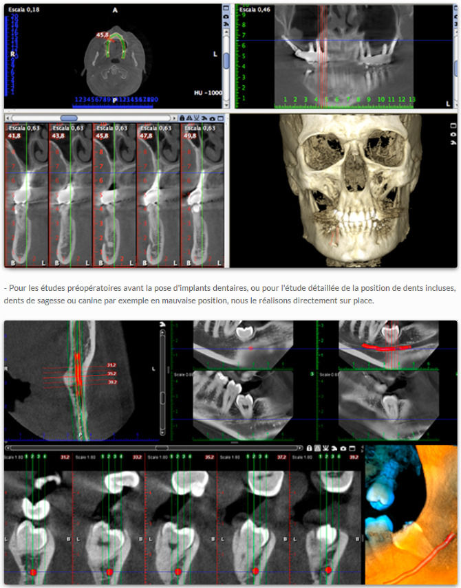
The extreme precision of the Cone Beam allows not only an in-depth examination of the skeletal state of the maxilla, but also the detection of anatomical elements such as the nerves and the mandibular canal. The latter extends from under the root of the tooth to the jawbone and contains nerve endings and arteries. Therefore, it is important to know the exact course so as not to touch it when inserting the dental implant.
In endodontics:
So-called “periodontal” x-rays help to ensure that the bone tissue and the subgingival part of the tooth are in good condition. However, a Cone Beam dental scan may be needed to check for progression of fractures (alveolar fractures or root fractures) infections or root canal healing. This allows doctors to assess the extent of the disease more accurately in case they need to determine the appropriate treatment later.
In surgery:
For example, the tooth may not fall out and get stuck in the jawbone. This is called an “impacted tooth” and may require surgery in some patients. In this context, Cone Beam dental scanners allow the practitioner to obtain more detailed information on the situation in order to guide him in the intervention process, to better plan the treatment, to carry out a complete infectious assessment and to identify very benign lesions.
Price of a Cone Beam exam?
The price of the dental scanner is indicated to you in full transparency before your visit. It depends on the area being studied and why it is required. We indicate in the quote the amount of the reimbursement made by the Health Insurance, so that you can benefit from all the information.
Why do a Cone Beam dental scanner?
The Cone Beam is:
- Taking shots in sections
- A high level of study precision
- Extensive observation of bone masses
However, Cone Beam has the advantage of being much faster and sharper when rotating for a few seconds. It analyzes small dental structures and identifies small elements such as nerves. By its action, it is possible to direct the rays to a specific area. In addition, this avoids irradiating parts of the skull that are not relevant to the examination process.
Thus, dental surgeons who use it expose their patients to a lesser amount of X-rays and thus have a high resolution image. They can thus establish their diagnosis, or plan the location of implants, for example.
It is obvious that if you suffer from periodontal disease, your dentist will have to determine the damage in order to propose an appropriate treatment. Then, Cone Beam lets him know exactly which areas are the most degraded.
A few tips:
In order to limit the appearance of dental problems, it is important to follow the rules ofgood oral hygiene:
- Brush your teeth at least twice a day with fluoride toothpaste
- Use dental floss or water flosser to complete your teeth brushing
- Make frequent appointments with your specialist
- Have a scaling done regular
For instance, in periodontal disease, the supporting tissues of the teeth are destroyed, which can lead to their loss. These fabrics are:
- The gums ;
- The nerves ;
- The blood vessels ;
- The alveolar bone.
Unfortunately, this situation cannot be changed. However, with prompt treatment, the disease can be slowed down or completely cured and the patient can retain tooth function. To prevent the appearance of this disease, descaling, mentioned above, is essential.
Indeed, as it develops on your teeth, dental plaque (a mixture of bacteria, saliva and food debris) gradually calcifies on contact with the minerals present in the mouth and turns into tartar. It is a hard, rough layer that clings to the teeth and serves as a vector for new diseases. If left unchecked, it builds up, thus peeling off the gum line and pushing down to the alveolar bone before attacking it.
If you have any questions or would like a quote, please do not hesitate to contact us!